. 2015 Mar 16;4:e04872. doi: 10.7554/eLife.04872

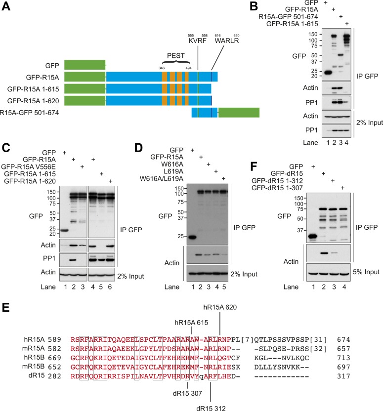
Figure 3. Actin associates with the conserved C-terminal portion of PPP1R15.

(A) Schematic diagram of human PPP1R15A (R15A) constructs used. Green indicates GFP. PEST repeats (between residues 346 and 494, orange), K555VRF558 (yellow), and W616ARLR620 (purple) sequences are identified. (B) Immunoblot for GFP, actin, and PP1 of HEK293T lysates from cells expressing indicated constructs and PP1, and subjected to GFP affinity purification (upper three panels). Immunoblot for actin and PP1 of 2% of input. (C) Immunoblot for GFP, actin, and PP1 of HEK293T lysates from cells expressing indicated constructs and PP1, and subjected to GFP affinity purification (upper three panels). Immunoblot for actin of 2% of input. (D) Immunoblot for GFP and actin of HEK293T lysates from cells expressing indicated constructs and subjected to GFP affinity purification (upper two panels). Immunoblot for actin of 5% of input (lower panel). (E) Sequence alignment of C-terminal portions of human (h) and murine PPP1R15A (mR15A) and PPP1R15B (mR15B) and Drosophila dPPP1R15 (dR15) with regions of homology boxed. Specific truncations are indicated. (F) Immunoblot for GFP and actin of HEK293T lysates from cells expressing indicated constructs and subjected to GFP affinity purification (upper two panels). Immunoblot for actin and PP1 of 2% of input.

DOI: http://dx.doi.org/10.7554/eLife.04872.007

Figure 3.

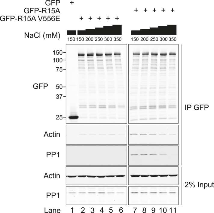
Figure 3—figure supplement 1. Immunoblot for GFP, actin, and PP1 of GFP-Trap pull-downs and 2% of input.

Figure 3—figure supplement 1.

HEK293T cells were transiently transfected with plasmids encoding the indicated constructs. After 36 hr, cells were lysed in GFP-Trap lysis buffer (150 mM NaCl, 10 mM Tris/Cl pH 7.5, 0.5 mM EDTA, 1 mM PMSF, and Protease Inhibitor Cocktail [Roche]) and post-nuclear supernatants were incubated with GFP-Trap beads at 4°C for 2 hr then washed four times in the same buffer. Next, samples were washed thrice in GFP-Trap lysis buffer supplemented with additional NaCl as indicated.
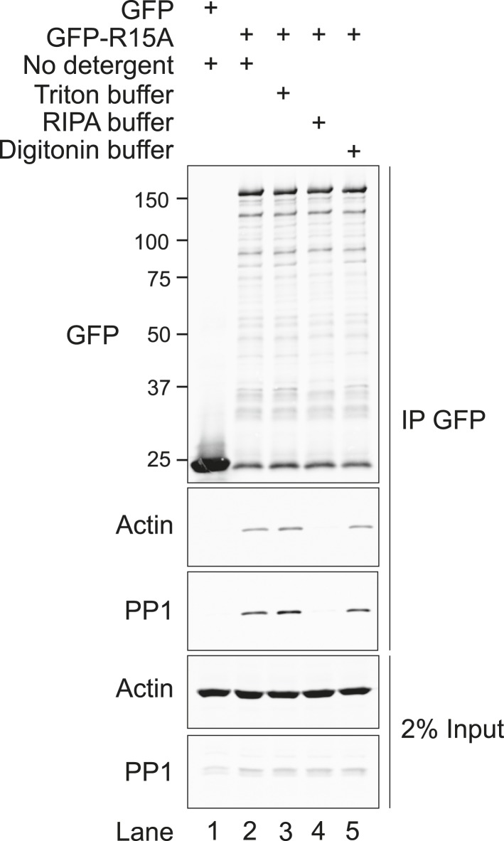
Figure 3—figure supplement 2. Immunoblot for GFP, actin, and PP1 of GFP-Trap pull-downs and 2% of input.

Figure 3—figure supplement 2.

HEK293T cells were transiently transfected with plasmids encoding the indicated constructs. After 36 hr, cells were lysed in GFP-Trap lysis buffer (150 mM NaCl, 10 mM Tris/Cl pH 7.5, 0.5 mM EDTA, 1 mM PMSF, and Protease Inhibitor Cocktail [Roche]) and post-nuclear supernatants were incubated with GFP-Trap beads at 4°C for 2 hr then washed once in the same buffer. Next, samples were washed thrice with no detergent (GFP-Trap lysis buffer), triton buffer (150 mM NaCl, 10 nM HEPES pH 7.4, 0.5% vol/vol triton X-100), RIPA buffer (150 mM NaCl, 50 mM Tris HCl pH 7.4, 1% vol/vol NP40, 0.5% vol/vol sodium deoxycholate, 0.1% vol/vol SDS) or digitonin buffer (150 mM NaCl, 50 mM Tris HCl pH 7.4, 0.1% vol/vol digitonin).