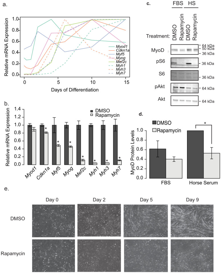
Figure 1. Rapamycin blocks C2C12 differentiation.
(a) The order of appearance of myotube differentiation markers over the course of 15 days in differentiation media only. This is representative of three independent experiments. (b) Differences in differentation marker transcripts when treated with DMSO (vehicle) or 500 nM rapamycin for 9 days, throughout the differentiation protocol. The most recent rapamycin administration was 1 day prior to cell lysis. These data represent the average of three wells, from a representative experiment (n = 3). Transcripts from both (a) and (b) were measured by qRT-PCR and normalized to Gapdh. (c) Representative western blot analysis of C2C12 cells treated for 4 h with Differentiation Media (2% Horse Serum; HS) or left in growth media (10% FBS) in the presence of 500 nM Rapamycin or DMSO. (d) Quantification of the blots in c (n = 4 independent experiments). Protein phosphorylation is presented as the intensity of the phosphospecific antibody band relative to that proteins total protein band. (e) Images of morphological changes in C2C12 myoblasts in response to 10 days of DMSO or rapamycin treatment througout the differentiation protocol (500 nM). Asterisks indicate p < 0.05. Data represents mean +/− standard error of the mean.

