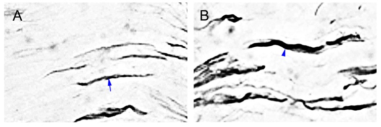
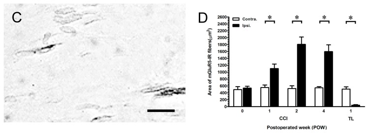
Figure 6.
Distribution of mGluR5-IR fibers along the distal compression stumps of sciatic nerve. The sciatic nerves at POW 1 from the ipsilateral sides of (A) sham-operated surgery, (B) CCI, and (C) TL were immunostained with antibody against mGluR5. (A) The mGluR5-IR fibers typically expressed the dense linear appearances (blue arrow); (B) The more thick forms were presented in mGluR5-IR fibers (blue arrowhead); (C) The mGluR5-IR fibers were shown with the weakened occurrences. Scale bar = 25 μm; and (D) The areas of GluR5-IR fibers were quantified as the mean ± SD (n = 5 at each time points after CCI, n = 5 at POW 1 after TL). Each bar of values showed in the contralateral sides (Contra., open bars) and ipsilateral sides (Ipsi., filled bars). Student’s t test was applied to examine the differences between the Contra. and Ipsi. at the same time points. * p < 0.05, indicated as a significant difference.


