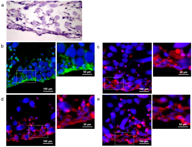
Figure 2.
DPSCs seeded onto hyaluronan-based scaffolds and cultivated with differentiation medium for 21 days. (a) H/E staining; Fluorescence images of cells expressing (b) collagen type I (green), a component of the extracellular matrix (ECM); (c) Von Willebrand factor (red), an endothelial marker; (d) Beta-III Tubulin (red), a neuronal marker; and (e) GFAP (red), a glial marker. Cell nuclei and the biomaterial are stained blue.

