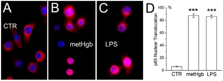
Figure 3.
metHgb causes NFκB activation in microglia. (A–C) Cultured microglia under control conditions (A); or after 60-min exposure to purified LPS-free metHgb (7 mg/mL) (B) or LPS (100 pg/mL) (C), immunolabeled with anti-p65 antibody; nuclei labeled with DAPI (blue); pink nuclei indicate nuclear translocation of p65; (D) Percent of nuclei showing translocation of p65 1 h after exposure to purified LPS-free metHgb (7 mg/mL) or LPS (100 pg/mL); 3 replicates per treatment; ***, p < 0.001.

