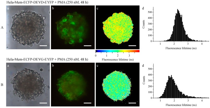
Figure 6.
Transillumination (a), fluorescence intensity (λ ≥ 515 nm); (b) and fluorescence lifetime (450–490 nm); (c) including histograms; (d) of HeLa cervical carcinoma cells expressing Mem-ECFP-DEVD-EYFP (A) or Mem-ECFP-DEVD-EYFP (B) after application of phorbol-12-myristate-13-acetate (PMA) (250 nM, 48 h). Light sheet based fluorescence microscopy of individual layers of a multicellular spheroid with 6–10 µm thickness each. Excitation wavelength: 391 nm. Scale bars: 50 µm.

