Figure 1.
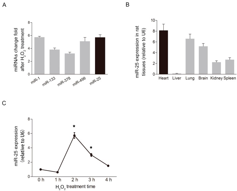
MiR-25 was elevated under oxidative stress. (A) MiR-25 expression was dramatically altered in response to oxidative stress compared to other miRNAs that are highly expressed in cardiac tissue (n = 3); (B) Quantitative RT-PCR confirmed the expression profile of miR-25 in different rat organs (n = 3); (C) Time course of the relative expression of miR-25 in cardiomyocytes in response to H2O2 stimulation (500 μM). The expression of miR-25 increased significantly at 2 h, and gradually returned to baseline by 4 h. Data are means ± SD from three independent experiments. * p < 0.05 vs. control (0 h).

