Figure 7.
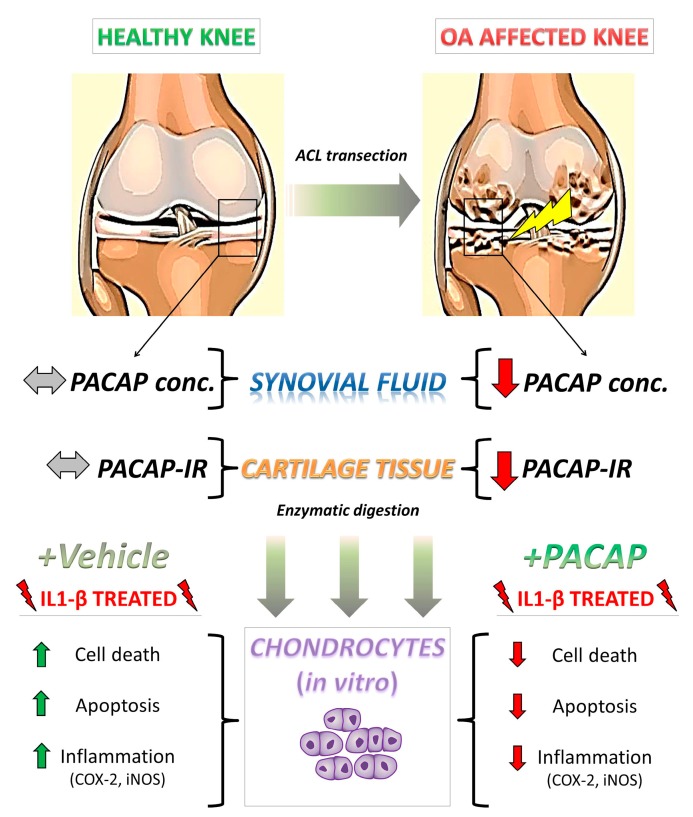
Schematic illustration depicting PACAP regulation in the proposed model of OA and its beneficial effects in isolated chondrocytes after IL-1β insult. Upon anterior cruciate ligament transection (ACLT), rats progressively develop clinical signs of OA, including cartilage deterioration and local inflammatory response [55,56]. In this scenario, PACAP immunoreactivity, which was detectable at high intensities in chondrocytes localized in the outer regions of the healthy cartilage is significantly reduced by experimental induction of OA. Such an event is accompanied by the reduction of PACAP concentration in the synovial fluid and a remarkable increase in intra-articular presence of the pro-inflammatory cytokine IL-1β, suggestive of an active inflammatory process. To assess whether PACAP contributed to prevent chondrocyte death, these cells were isolated through enzymatic digestion and challenged with IL-1β, to partly mimic the inflammatory milieu observed in vivo. Results show that PACAP treatment ameliorated most of the detrimental effects of the cytokine, acting both as a chondroprotective and anti-inflammatory molecule. PACAP conc. = PACAP concentration; PACAP-IR = PACAP immunoreactivity.

