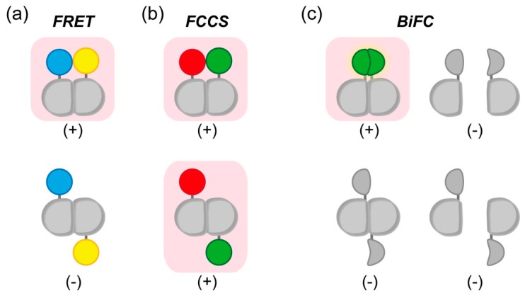
Figure 4.
Detection of orientation-dependence or -independence of dimer formation by FRET, FCCS, or BiFC. Magenta background color and (+)/(−) indicates signal emission in each methodology; (a) Restriction of FRET effect in dimerization depending on the tagging position of fluorescent molecule. Blue and yellow colors indicate FRET donor and acceptor, respectively; (b) Despite the tagging positions of the fluorophores, FCCS can detect dimerization. Green and red colors indicate fluorescence tag; (c) In addition to monomeric state (right top and bottom) with no fluorescence, not-appropriate tagging position of the fragments also shows no fluorescence (left bottom). In BiFC, as in FRET, only the appropriate positioning of the protein domains results in fluorescence (green color, left top).

