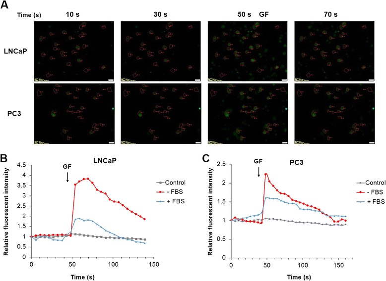
Figure 6.

Effects of GF on cytosolic Ca2+ signal. (A) Fluorescent Images for LNCaP and PC3 cells treated with GF. ROI 1–20 were selected cells; ROI 21–23 were selected as background. Fluorescent intensity was quantified by background subtracted and normalized from the start. Cells were stained with Fluo-4-AM, and images were acquired by live cell imaging system (Ex: 488 nm, Em: 543 nm BP), for 3 min with 5 sec interval. GF was added at 50 sec and the arrows showed the addition of 10 μM GF. (B) Time-response curve (mean) was determined from data generated in selected 20 LNCaP cells; (C) Time-response curve (mean) was determined from data generated in selected 20 PC3 cells. Experiments were repeated 3 times.
