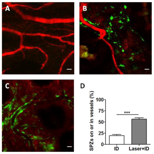
Figure 3.
Confocal microscopy of sporozoites in the skin. (A) Blood vessels were marked by Texas red-dextran (MW 70,000). (B and C) Representative images of CFSE-stained sporozoites in un-treated (B) or laser-treated skin (C). Bar =10 μm. (D) Percentages of sporozoites in association with vessel walls or inside the vessels. Data are shown as means ± SD. n=10, ***p<0.001.

