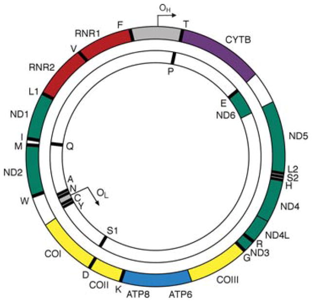
Figure 1.
A schematic of the 16.6 kb circular, double-stranded human mitochondrial DNA molecule. The outer circle represents the heavy (H) strand of mtDNA and the inner circle the light (L) strand. OH and OL are the origins heavy-strand and light strand and replication. The gray region represents the mitochondrial control region also known as the D-loop. Genes encoding subunits of respiratory chain complex I are shown in green, the MTCYB gene of complex III in purple, the three catalytic subunits of complex IV in yellow and MTATP6 and MTATP8 of complex V in blue. The two ribosomal RNAs are shown in red and the 22 transfer RNAs represented as black bars and denoted by their single-letter abbreviations.

