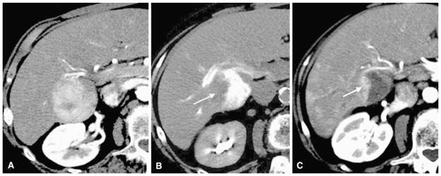
Fig. 5.

An 84-year-old woman with hepatocellular carcinoma. A Arterial phase preprocedure contrast-enhanced CT shows index tumor in segments V and VI. B Immediate postembolization CT shows DCR on the right side of the tumor (arrow) despite grade 2 TCR and grade 2 VCR. C Follow-up contrast-enhanced CT shows partial residual enhancing tumor morphology (arrow) corresponding to the contrast retention defect
