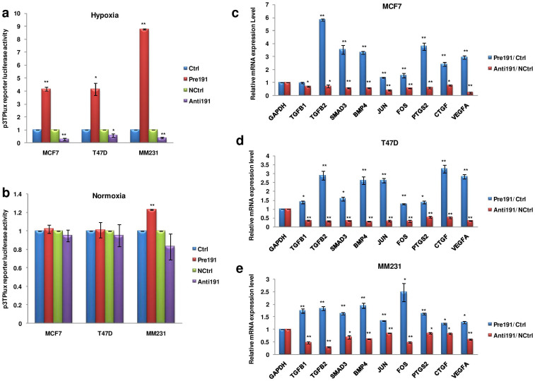
Figure 4. miR-191 promotes TGFβ-signaling in hypoxic microenvironment.
(a, b). Bar graph represents relative luciferase activity of p3TP-Lux reporter plasmid on overexpression/inhibition of miR-191 under hypoxic (a) and normoxic conditions (b). (c–e). qRT-PCR data showing expression level of genes belonging to TGFβ-pathway in response to miR-191 overexpression/inhibition in hypoxic microenvironment in a panel of breast cancer cell lines- MCF7 (c), T47D (d), MM231 (e). The graphical data points in a-e represent mean ± S.D of at least three independent experiments. (*P < 0.05, **P < 0.01). Error bars denote ± SD.

