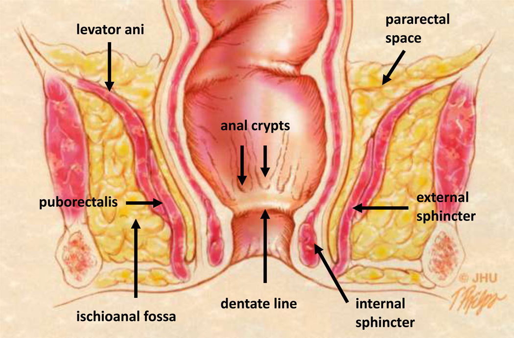
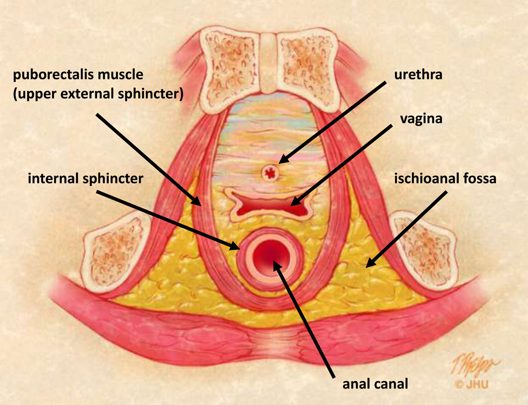
Fig 2.
Pelvic floor and anal canal anatomy.
2A – Coronal anatomic view of the pelvis. The anal glands are the starting location for cryptoglandular disease and emerge in the anal crypts along the dentate line. The interstitial tissue between the internal and external sphincters allows access to the pelvic compartment and pararectal space above the levator ani. The puborectalis muscle forms the superior aspect of the external sphincter and is functionally part of the levator ani muscle complex (17).
2B – Axial anatomic view of the female pelvis and uppermost anal canal mechanism. Cryptoglandular disease can spread circumferentially in the interstitial tissue between the internal and external sphincter. The fat filled ischioanal fossa can also become infected once both sphincters are breached.


