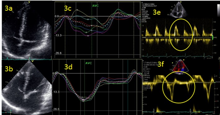
Figure 3.

(a, b) Left atrium (LA) follow-up 2 years of triathlete with enlargement of LA and atrial fibrillation events. (b, c) Strain curves of left ventricle during atrial fibrillation (c) and 24 h after recovery in sinus rhythm (d). (e, f) Doppler measurement of diastolic function: conventional (e) and using tissue Doppler imaging (TDI).
