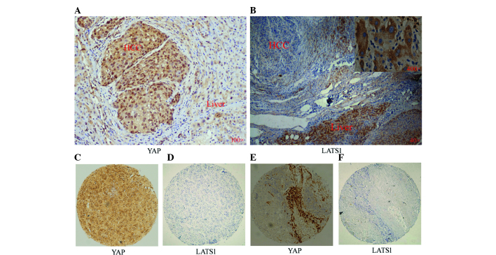
Figure 1.
Expression of YAP and LATS1 in HCC and PCT tissues. Representative images of (A) YAP staining (magnification, ×100) or (B) LATS1 staining in HCC (magnification, ×40) and PCT (magnification, ×40 and ×400) as detected by immunohistochemistry. (C and D) Identical HCC tissue slice stained for YAP and LATSI, respectively. (E and F) Another identical HCC tissue slice stained for YAP and LATSI, respectively (magnification, ×40). YAP, yes-associated protein; LATS, large tumor suppressor 1; HCC, hepatocellular carcinoma; PCT, para-cancerous tissue.

