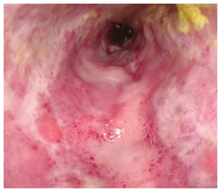
Figure 2.
A 38-year old woman with history of an unknown connective tissue disorder who presents with ischemic colitis secondary to inferior mesenteric arteriovenous malformation.
FINDINGS: Photograph of the sigmoid colon obtained during flexible sigmoidoscopy demonstrates diffuse ulceration, edema, and exudate with luminal narrowing. The mucosa does not appear necrotic.
TECHNIQUE: Flexible sigmoidoscopy.

