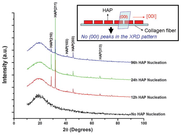
Figure 3.
XRD patterns of SIS membranes mineralized for 0, 12, 24 and 96 h. The inset schematically illustrates that when HAP has c-axis preferred orientation along the collagen fibers lying on the SIS membrane, the (00l) peaks will be absent in the XRD patterns, which is the actual case in the XRD patterns shown here.

