Figure 2.

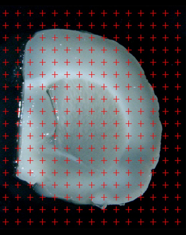
The slab is photographed under an anatomical microscope at a magnification of 10×. A point grid is randomly superimposed on the photographed slab, and the points that touch the cortex are counted.

The slab is photographed under an anatomical microscope at a magnification of 10×. A point grid is randomly superimposed on the photographed slab, and the points that touch the cortex are counted.