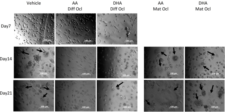
Fig 3. Effects of AA and DHA on osteoclast cell morphology.
Osteoclast differentiation was stimulated in CD14+ monocytes by the addition of 25 ng ml-1 M-CSF and 30 ng ml-1 RANKL as described in Materials and Methods. Vehicle (0.08% ethanol) or LCPUFAs (40 μM) were added from day 3 in differentiating osteoclasts and from the onset of resorption (day 12–14) in mature osteoclasts. Osteoclasts were visualized under a microscope and PlasDIC images were taken throughout the culture period. Large multinucleated osteoclasts are indicated with black arrows. The results are representative of three independent experiments conducted in triplicate. Scale bar = 100 μm. Diff Ocl—differentiating osteoclasts. Mat Ocl—mature osteoclasts.

