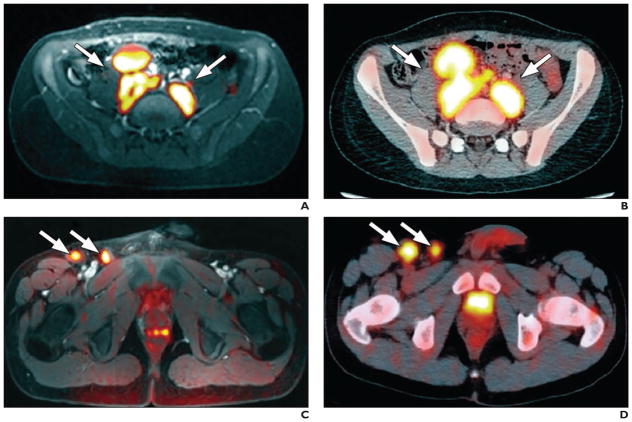
Fig. 1. Hodgkin lymphoma. (Reprinted with permission from [11]).
A and B, Whole-body diffusion-weighted MR image with ferumoxytol contrast administration (A) and FDG PET/CT image (B) in 15-year-old patient show positive retroperitoneal involvement (arrows) of stage IIIA Hodgkin lymphoma.
C and D, Whole-body diffusion-weighted MR image with ferumoxytol contrast administration (C) and FDG PET/CT image (D) in 14-year-old patient with stage IIB Hodgkin lymphoma show positive right inguinal lymph nodes (arrows).

