FIGURE 1:

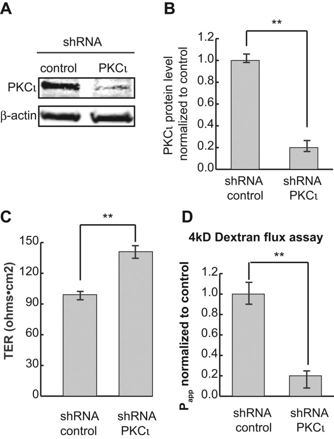
PKCι knockdown decreases the permeability of MDCK II monolayers. (A) PKCι was knocked down by lentiviral shRNA, and cell lysates were analyzed by Western blotting. β-Actin was used as loading control. (B) Quantification of protein levels in A. (C) TER measurement shows PKCι reduction results in a slight but significant increase of TER. (D) The 4- kDa FL-dextran flux assay shows that knockdown of PKCι significantly decreases epithelial permeability. Each experiment was performed in triplicate and repeated at least three times. Error bars represent SEM. **p < 0.001.
