Fig 2.

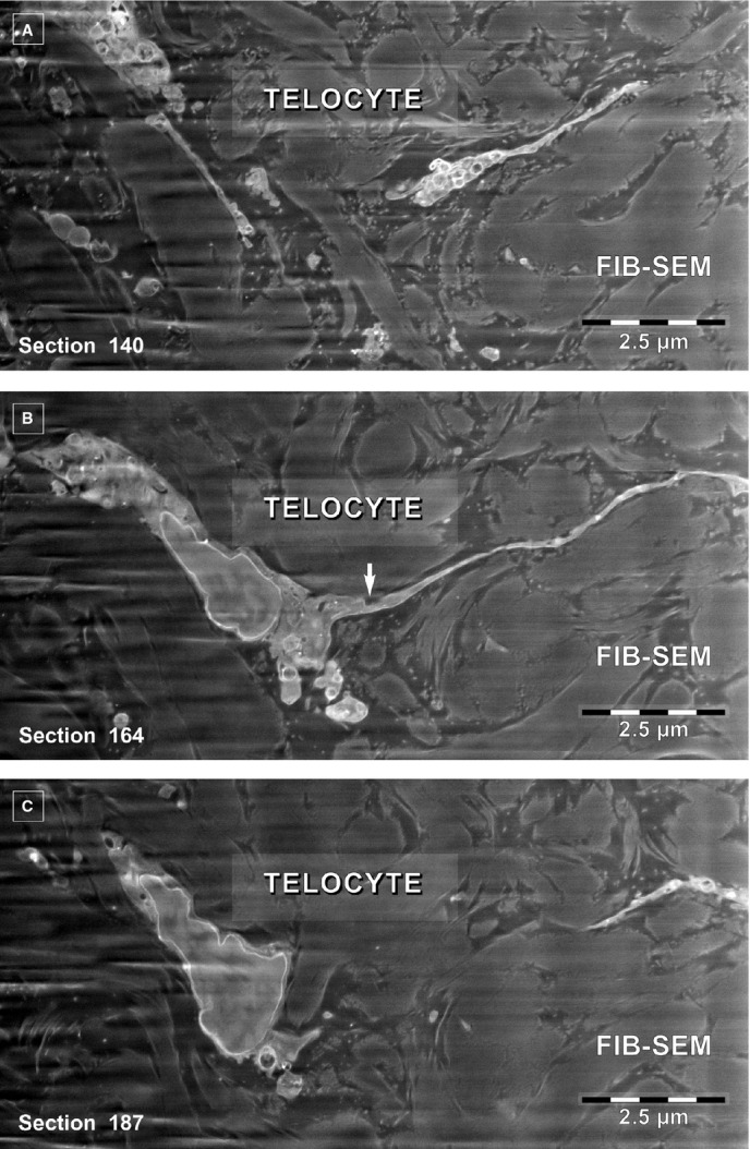
(A–C) FIB-SEM backscattered electron images. Three non-consecutive serial images obtained at ∼1.2 μm z-interval show the narrow emergence (arrow) of a telopode from the cellular body of a telocyte.

(A–C) FIB-SEM backscattered electron images. Three non-consecutive serial images obtained at ∼1.2 μm z-interval show the narrow emergence (arrow) of a telopode from the cellular body of a telocyte.