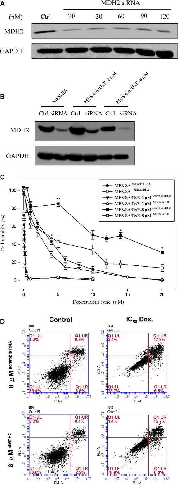
Fig 7.

Effect of doxorubicin on cell viability of MDH2 siRNA-silenced MES-SA, MES-SA/Dx-2 μM and MES-SA/Dx-8 μM cells. (A) Efficiency of MDH2 siRNA on the inhibition of MDH2 expression in MES-SA/Dx-8 μM cells. MES-SA/Dx-8 μM cells grown overnight were treated with indicated concentrations of MDH2-specific siRNA for 24 hrs. Expression of MDH2 in MES-SA/Dx-8 μM cells were monitored with immunoblotting by using primary antibodies against MDH2. (B) MES-SA, MES-SA/Dx-2 μM and MES-SA/Dx-8 μM cells grown overnight were pre-treated with 20 nM MDH2-specific siRNA or scramble siRNA with similar GC content. Expression of MDH2 in MES-SA, MES-SA/Dx-2 μM and MES-SA/Dx-8 μM cells were monitored by immunoblotting by using primary antibodies against MDH2. (C) MTT-based viability assays were performed where 5000 MES-SA, MES-SA/Dx-2 μM and MES-SA/Dx-8 μM cells seeded into 96-well plate for overnight incubation followed by pre-treated with 20 nM MDH2-specific siRNA combining with corresponding scramble siRNA. After 24 hrs, cells were treated indicated concentrations of doxorubicin for 24 hrs followed by incubated with MTT and then DMSO added and the plates shaken for 20 min. followed by measurement of the absorbance at 540 nm. Values were normalized against the untreated samples and are the average of 4 independent measurements ± SD. (D) MES-SA/Dx-8 μM cells were treated with IC50 concentrations of doxorubicin or left untreated for 48 hrs. After treatment, 106 cells were incubated with Alexa Fluor 488 and propidium iodide (PI) in 1× binding buffer at room temperature for 15 min., and then stained cells were analysed by flow cytometry to examine effect of doxorubicin on apoptosis in MES-SA/Dx-8 μM cells and MDH2 siRNA silenced MES-SA/Dx-8 μM cells. Annexin V is presented in x-axis as FL1-H, and PI is presented in y-axis as FL2-H. LR quadrant indicates the percentage of early apoptotic cells (Annexin V positive cells), and UR quadrant indicates the percentage of late apoptotic cells (Annexin V positive and PI positive cells).
