Fig 2.

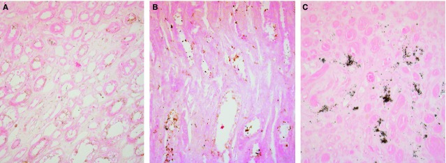
von Kossa staining of control papillary tissues. Light microscopy images (200×) of von Kossa-stained sections of control (A and B) and MSK (C) renal papillary interstitium. No calcium is visible in control tissues.

von Kossa staining of control papillary tissues. Light microscopy images (200×) of von Kossa-stained sections of control (A and B) and MSK (C) renal papillary interstitium. No calcium is visible in control tissues.