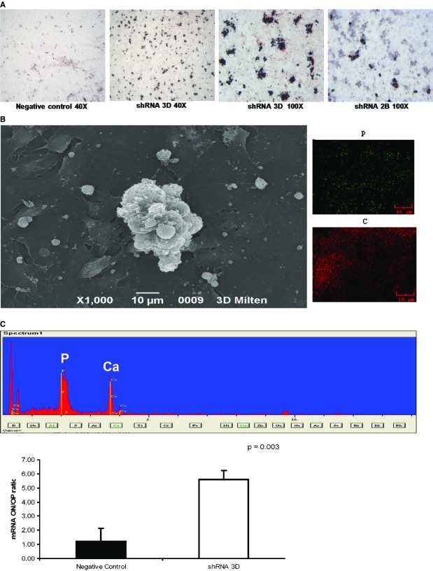
Fig 11.
Detection of Ca2PO4 deposits in HK2 cells cultured in osteogenic medium. (A) Light microscopy images (40× and 100×) of von Kossa staining reveal calcium deposits in cell aggregates. The low magnification image reveals that the dark deposits were much more abundant in the silenced clones than in the control clones. The figures are representative of the results of three experiments. (B) SEM images and spectrum confirming Ca2PO4 deposition. Calcium (green dots) and phosphorus (red dots) were colocalized. (C) Real-time PCR results for the ratios of osteonectin to osteopontin mRNAs, indicating that the balance between pro- and anti-osteogenic factors in the silenced clones was tipped in favour of an osteogenic process. The results represent the mean ± SD of three separate experiments performed in triplicate.

