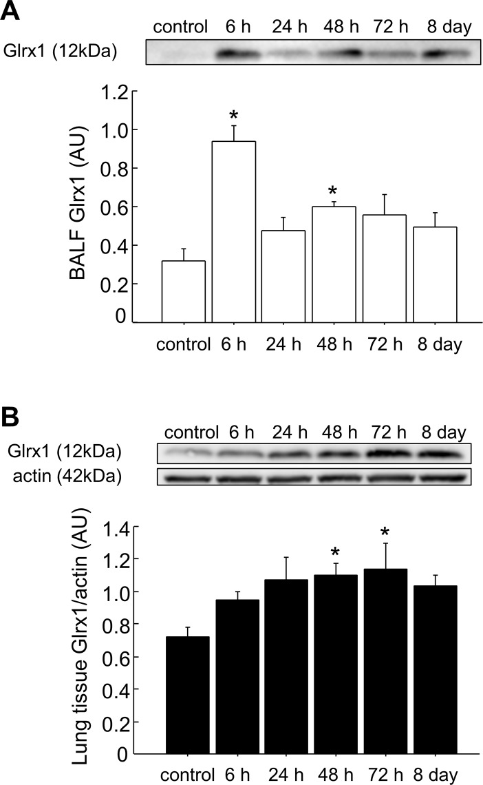
Fig 1. Expression of Glrx1 in the BALF and lung tissues of mice after OVA challenges.
BALF (A) and lung homogenates (B) from OVA-challenged mice were analyzed by western blot analysis for Glrx1 expression at the indicated time points (6, 24, 48, and 72 h, and 8 days after the last challenge with OVA). Actin was used as a loading control for lung homogenates; data are presented as means ± SEM. *p < 0.05 was considered significant in the comparisons with control mice.

