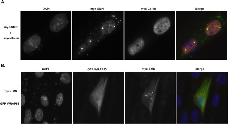
Fig 5. Co-expression of SMN and coilin in the WI-38 primary cell line promotes foci formation.
(A) WI-38 cells were co-transfected with myc-tagged SMN and coilin, followed by fixation and detection of the expressed proteins using anti-SMN or anti-coilin antibodies and appropriate secondary antibodies. DAPI was used to stain the nucleus. In the merged image, the nucleus is blue, SMN is green and coilin signal is red. Foci with co-localized SMN and coilin are yellow. (B) WI-38 cells were co-transfected with myc-SMN and GFP-WRAP53, followed by fixation and detection of the expressed proteins. Anti-SMN antibody was used to detect SMN and the nucleus was stained with DAPI. In the merged image, SMN is red, GFP-WRAP53 is green and the nucleus is blue.

