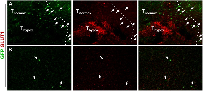
Fig 3. GFP positive pericytes are found preferably within hypoxic regions of glioma.
(A) GFP positive cells (arrows) are attracted more numerous within the penumbra zone around GLUT1 positive hypoxic regions close to the GL261 tumor border (dashed), scale bar is 200 μm. Normoxic and hypoxic parts of the tumor are marked with Tnormox and Thypox, respectively. (B) Few GFP positive pericytes (arrows) are found at normoxic regions within the tumor, scale bar as in A.

