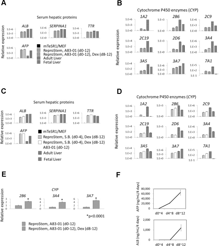
Fig 2. Hepatic differentiation of hiHSCs by depletion of FGF-2.
Self-renewing hiHSCs, clones AFB1-1 and NGC1-1, differentiate into hepatocyte-like cells with the omission of FGF-2 from ReproStem medium. (A–E) Gene expression was analyzed by quantitative RT-PCR at day 12. Gene symbols are shown for serum hepatic proteins and cytochrome P450 enzymes. The terms of additions are indicated in parentheses. (A–D) Relative expression is shown with a logarithmic axis histogram. The expression is normalized to 1 in the self-renewing hiHSCs (mTeSR1/MEF) and compared to that of hepatocyte-like cells. Total RNAs of human fetal and adult livers were utilized as robust controls. Data are presented as mean+SEM and represent a minimum of three independent samples with at least two technical duplicates. (A, B) Clone AFB1-1 differentiated in the medium including an inhibitor (0.5 μM A83-01) of TGF-β signaling or the medium including 0.5 μM A83-01 plus 0.5 μM dexamethasone (Dex). See also S11 and S12 Figs. (C, D) Clone NGC1-1 differentiated in the medium including 0.5 μM sodium butyrate (S.B.) plus 0.5 μM Dex or the medium including 0.5 μM S.B. plus 0.5 μM Dex plus 0.5 μM A83-01. See also S13 and S14 Figs. (E) Clone AFB1-1 differentiated in the medium including 0.5 μM A83-01 or the medium including 0.5 μM A83-01 plus 0.5 μM Dex. Relative expression is normalized to 1 in the medium without Dex and is shown in the histogram. The expressions of cytochrome P450 enzymes (CYP2B6, 3A4, and 3A7) are induced by the addition of Dex. Asterisk indicates statistical significance as determined by t test. *p < 0.0001. (F) Release of human ALB and AFP was measured by ELISA on samples (supernatants of clone AFB1-1 differentiated with the medium including 0.5 μM A83-01) at three time points. Data are presented as mean±SEM and represent a minimum of three independent samples with at least two technical duplicates. See also Table 1.

