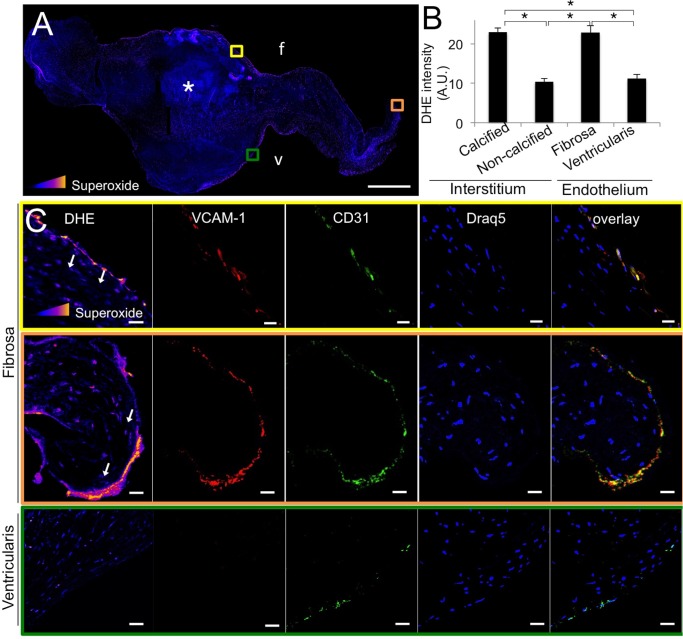
Fig 1. Superoxide is elevated in the endothelium of calcified human aortic valves.
A, Superoxide staining (DHE) of calcified human aortic valve leaflets. Asterisks indicate calcific nodule. Intensity of superoxide staining is colorimetrically scaled, with yellow indicating most intense (inset triangle). Colored boxes indicate region magnified in lower panels; f indicates fibrosa, v indicates ventricularis. Scale bar = 1 mm. B, Quantification of DHE intensity (superoxide) across all valve samples, presented as DHE intensity in different regions of calcified valves (cHAV), n = 21. * indicates p < 0.05 between indicated groups. C, Co-localization of elevated superoxide with VCAM-1 and CD31 expression in calcified human aortic valve leaflet endothelium. Representative images from n = 21 valves. Scale bar = 20μm.

