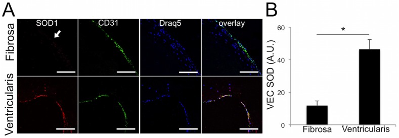
Fig 2. Calcified human aortic valves have lower expression of SOD on the fibrosa endothelium than on the ventricularis.

A. Immunofluorescence for endothelial protein CD31 and SOD1 revealed little to no expression of SOD1 (arrow) on the fibrosa endothelium of calcified aortic valve leaflets. The ventricularis showed strong SOD1 expression in the endothelium. B, Quantification of the pixel intensity of SOD1 in the CD31+ endothelial cells in cHAV, n = 21. The fibrosa endothelium had significantly less SOD1 than the ventricularis in cHAV. * indicates p < 0.05 between groups, Student’s t-test.
