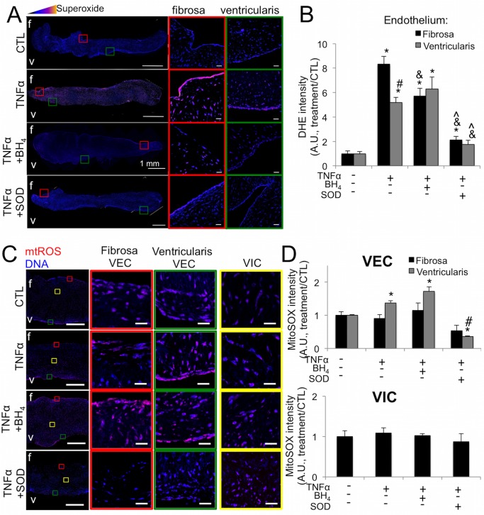
Fig 5. TNFα induces side-specific endothelial oxidative stress in ex vivo aortic valve leaflets.
A, TNFα causes increased superoxide in the fibrosa endothelium of ex vivo porcine aortic valve leaflets cultured for 21 days, revealed by DHE staining. Superoxide is mitigated on the fibrosa by co-treatment with BH4 and on both the fibrosa and ventricularis by peg-SOD. Pixel intensity of superoxide levels is scaled colorimetrically (inset triangle) to show regions of highest superoxide. Colored boxes indicate regions magnified on right, showing endothelium on fibrosa and ventricularis sides of valve. Scale bar is 1mm in left images, 20μm in magnified images. B, Quantification of endothelial superoxide in ex vivo aortic valves stained with DHE reveals side-specific rescue-effects of BH4 and more pronounced mitigation of superoxide on both sides of the valve by peg-SOD. C, TNFα increases mtROS in the ventricularis VEC of ex vivo AV leaflets. D, Quantification of mtROS fluorescenc. Ventricularis-specific increases in mtROS are mitigated by peg-SOD but not BH4. * indicates p < 0.05 versus control condition. # indicates p < 0.05 versus fibrosa endothelium in same condition. & indicates p < 0.05 versus same side endothelium in TNFα condition. ^ indicates p < 0.05 versus same side endothelium in TNFα+BH4. N = 6.

