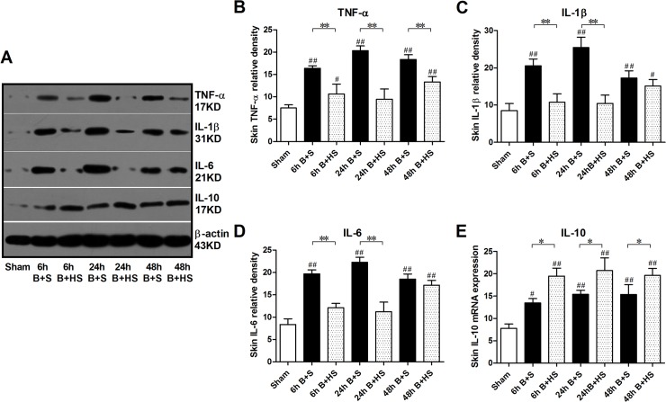
Fig 4. Western blot analysis of inflammatory cytokines in wound interspaces after burn with HS treatment.
Parallel increases in TNF-α, IL-1β, and IL-6 protein expression (as proinflammatory cytokines) were found at the three time points via western blot, while IL-10 also increased after burn to mediate the anti-inflammatory response. HS administration reduced the elevations of the three selected pro-inflammatory cytokines, and it further raised the levels of IL-10 to enhance the anti-inflammatory effect in wound tissues. The sample size was n = 6 for each group. The results were expressed as the mean±SEM. *p<0.05, **p<0.01, versus burn+saline; #p<0.05, ##p<0.01, versus sham. B+S, burn+saline; B+HS, burn+hydrogen-rich saline.

