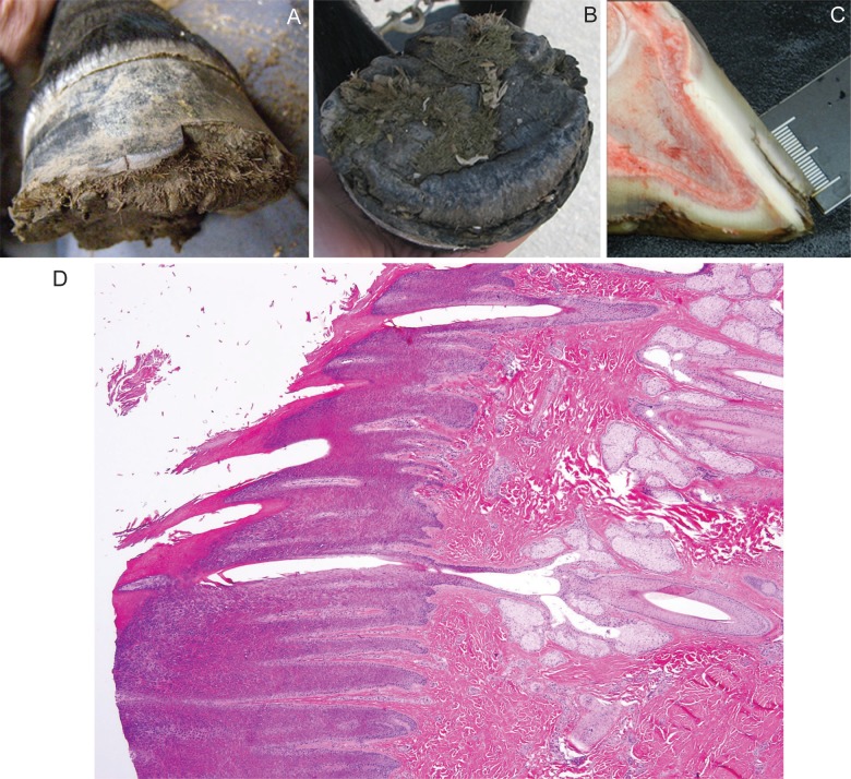
Fig 1. Hoof from a Connemara pony foal affected with hoof wall separation disease (HWSD).
(A) Note the dorsal hoof wall separation at the sole and (1B) proliferative horn on the solar aspect of the hoof (1B). (1C) A sagittal section of a post-mortem HWSD-affected hoof demonstrates that the dorsal hoof wall fissure occurs outside of the white line. (1D) Coronary band, periople (hematoxylin & eosine stain): normal transition from haired skin into periople is evident.

