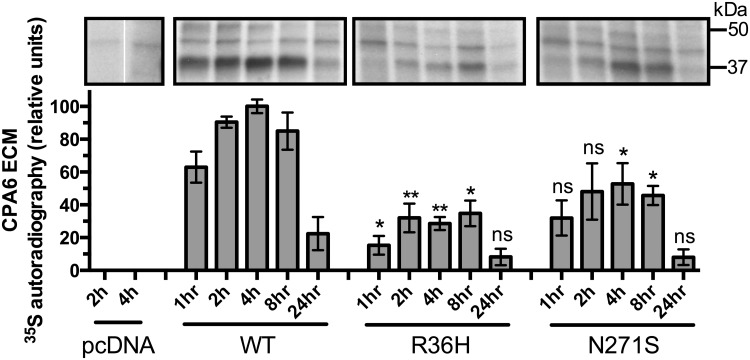
Fig 6. Pulse-chase analysis of HEK293T cells transfected with WT or mutant proCPA6.
Cells were labeled with 35S methionine and cysteine for 20 minutes (pulse), washed once, and incubated for the time indicated (chase) in media containing non-radioactive methionine and cysteine. ECM was extracted with SDS and analyzed on a denaturing polyacrylamide gel, which was dried and exposed to film for 7–10 days. CPA6, which runs ~37 kDa, was not detected in the cells transfected with pcDNA vector alone. Arg36His samples showed reduced levels of CPA6 relative to WT at all time points, which was statistically significant at all time points except 24h. Asn271Ser samples showed a trend towards reduced levels of CPA6 at all time points, with significant reductions in CPA6 levels at the 4 and 8 hour time points. Comparisons were performed across CPA6 variants for each time point; ns, not significant; *, p<0.05; **, p<0.01 relative to the same time point for the WT sample. Error bars show standard error of the mean (n = 3).

