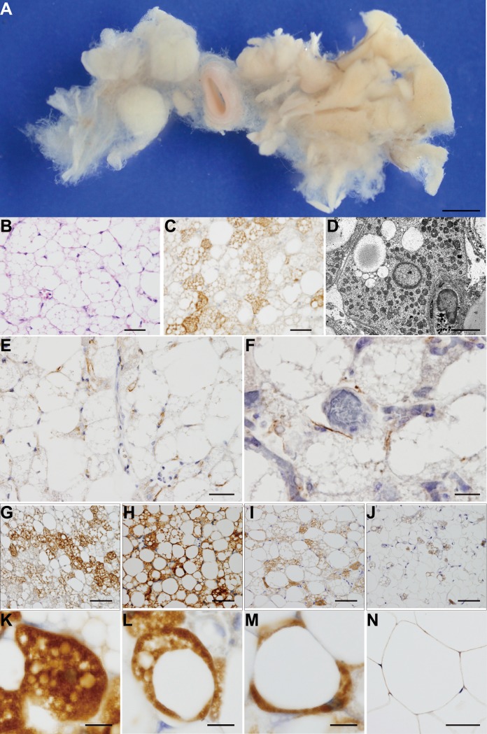
Fig 1. Overview of BATs and cells.
(A) Typical BATs in the thoracic periaortic area of a 16-year-old male. (B) HE staining, (C) UCP1 immunohistochemical staining and (D) transmission electron microscopy. Immunohistochemical staining for TH (E) and vAChT (F). Morphological properties of UCP1 immunohistochemical staining of BATs for the multilocular (K), paucilocular (L), and unilocular (M) types. (G) Multilocular-predominant, (H) paucilocular-predominant, (I) paucilocular-predominant, (J) unilocular-predominant. (N) The unilocular adipocytes of subcutaneous white adipose tissue. Scale bars: (A) 10 mm, (B)(C)(E)(F)(G)(H)(I)(J)(N) 50 μm, (D) 5 μm, and (K)(L)(M) 10 μm.

