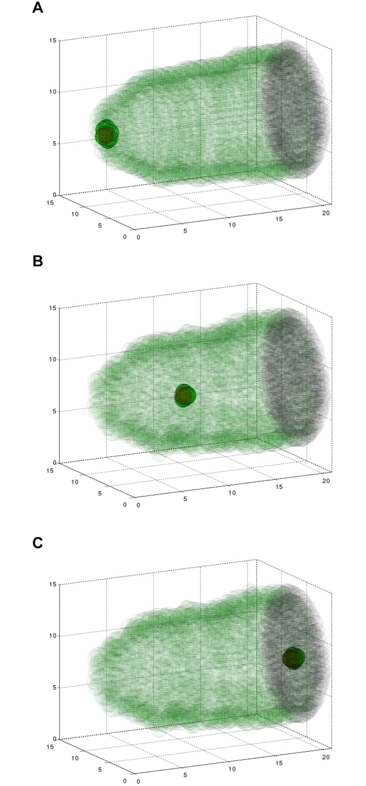
Fig 3. Simulating the three dimensional model results in collective migration.
A simulation showing six border cells (green), two polar cells (red), the epithelium (transparent green), and the surface of the oocyte (black, right) at three time points during the migration. Fifteen nurse cells are situated inside the egg chamber, but are not plotted so as to maintain clarity of this three dimensional structure. Polar cells are surrounded by border cells, making them hard to distinguish. (A) At 2 minutes, cells are beginning to invade between nurse cells. (B) At 2.4 hours, the cluster is about halfway to its destination. (C) At 5.6 hours, the border cell cluster has reached the edge of the oocyte. See also Supplemental S3 Movie.

