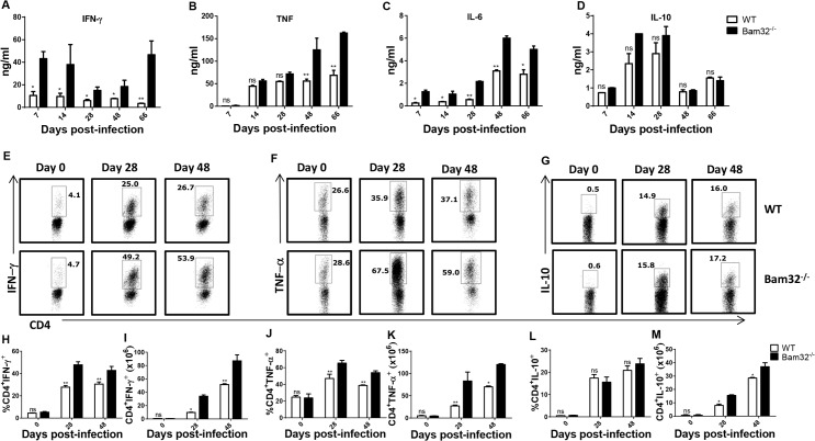
Fig 2. Increased production of proinflammatory cytokines by T. congolense infected Bam32-/- mice.
WT and Bam32-/- mice were infected with 103 T. congolense. At indicated times, infected mice were sacrificed, the spleen cells were cultured for 72 hr and the culture supernatant fluids were assayed for IFN-γ (A), TNF-α (B), IL-6 (C) and IL-10 (D) by ELISA. Some spleen cells were also directly stimulated ex-vivo with PMA, BFA and ionomycin for 3–5 hr, stained for intracellular expression of IFN-γ (E, H and I), TNF-α (F, J and K) and IL-10 (G, L and M), and analyzed by flow cytometry. Fig 2H–2M are bar graphs representing the means +/- standard error of the percentages (H, J and L) and absolute numbers (I, K and M) of CD4+ T cells that express IFN-γ (H and I), TNF-α (J and K) and IL-10 (L and M), respectively. Results are representative of 2 different experiments (n = 3–5 mice per experiment) with similar outcome. ns, not significant; *, p < 0.05; **, p < 0.01.

