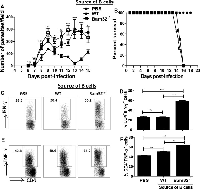
Fig 6. Bam32-/- B cells do not mediate parasite control in B cell deficient mice.
B cells were isolated from naïve WT and Bam32-/- mice and adoptively transferred intravenously into μMT mice. Control mice received PBS. Forty-eight hours after transfer, recipient mice were infected with 103 T. congolense, parasitemia (A) and survival (B) were monitored. At sacrifice, spleen cells were stimulated directly ex-vivo with PMA, BFA and ionomycin for 3–5 hr, stained and assessed for intracellular cytokine expression by flow cytometry. Shown are representative dot plots (C and E) and bar graphs (D and F) showing the means +/- standard error of the percentages of CD4+ T cells that express IFN-γ (C and D) and TNF-α (E and F). Data shown are representative of 2 separate experiments (n = 3 mice per experiment) with similar outcome. *, p <0.05; **, p < 0.01; ***, p < 0.001; ns, not significant.

