Figure 2.
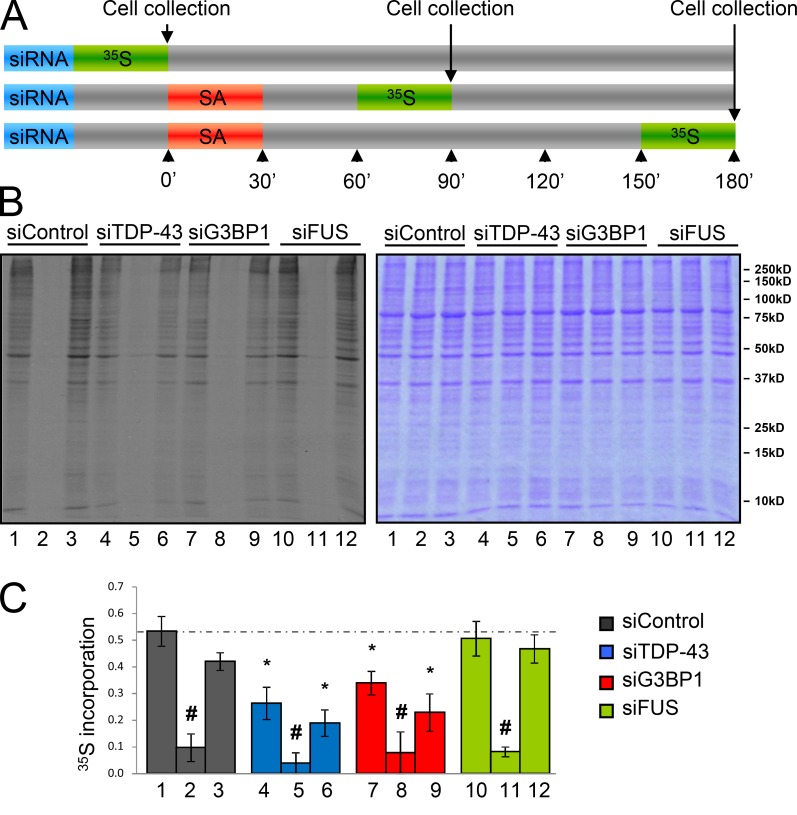
TDP-43 and G3BP1 depletion induce a defect in basal translation. (A–C) HeLa cells were transfected with the indicated siRNAs for 72 h. Cells were stressed with 0.5 mM SA for 30 min and collected at 90 (lanes 2, 5, 8, and 11) and 180 min (lanes 3, 6, 9, and 12) and before stress (lanes 1, 4, 7, and 10). All samples were incubated with 50 µM [35S]methionine 30 min before cell lysis. Samples were prepared in Laemmli buffer and subjected to SDS-PAGE. (A) Experimental design. (B) Autoradiography of the cell lysates (left) and same gel stained with Coomassie blue to demonstrate equivalent protein loading (right). (C) The histogram shows densitometric quantification of the entire lane expressed relative to the Coomassie-stained gel. The means ± SEM (error bars) of ≥3 independent experiments are plotted. *, P < 0.05, compared with siControl; #, P < 0.05, compared with the unstressed sample.

