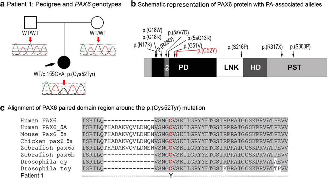
Fig. 1.
a Patient 1: Pedigree and PAX6 genotypes. DNA chromatograms of the proband’s and parental PAX6 sequences are shown with a position of c.155G>A, p.(Cys52Tyr) mutation indicated with red arrows; proband is designated with a black arrow. b Schematic representation of the PAX6 protein with PA-associated alleles. PD Paired domain, LNK linker region, HD homeodomain, PST transactivation domain rich of proline, serine, and threonine, 5a- 14-a.a. encoded by the alternatively spliced exon 5a. Positions of all previously reported PAX6 mutations associated with Peters anomaly are shown with black arrows; the cysteine residue at position 52 is shown in red font. c Alignment of PAX6 paired domain region around the p.(Cys52Tyr) mutation. Human PAX6 (NP_000271.1), human PAX6-5A (NP_001245391), mouse Pax6-5a (NP_001231127), chicken pax6-5a (NP_990397), zebrafish pax6a (NP_571379), zebrafish pax6b (NP_571716), Drosophila ey (NP_001014693) and Drosophila toy (NP_524638) sequences were used for the alignment; the position of p.(C52Y) is shown with red arrow

