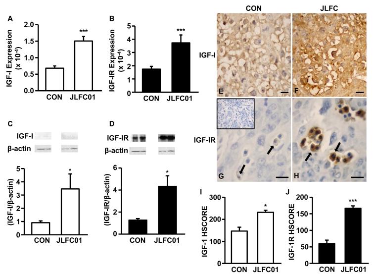
Figure 6. LFC01 increases IGF-I and IGF-1R expression in the placenta of CBA/J mice.
Placental IGF-I and IGF-IR expression was evaluated by (A, B) qRT-PCR, (C, D) Western blot with densitometry. The expression of IGF-I and IGF-IR in placentae from CBA/J mice treated in the absence (E, G) or presence (F, H) of JLFC01 was evaluated by immunohistochemistry (400x). IGF-I and IGF-IR immunoreactivity was semi-quantitatively evaluated using the following intensity categories: 0, no staining; +, weak but detectable staining; ++, moderate or distinct staining; and +++, intense staining. (I, J) A histological score (HSCORE) was calculated using the formula HSCORE = ∑(Pi × i), where i represents the intensity scores, and Pi is the corresponding percentage of the cells. Five fields/slide were evaluated by 2 investigators blinded to the tissue source. The results of densitometry and immunohistochemistry are reported as mean ± SEM. n=3; * p < 0.05; *** p < 0.005. Inset: IgG control; Scale bar: 50 μm.

