Figure 6.
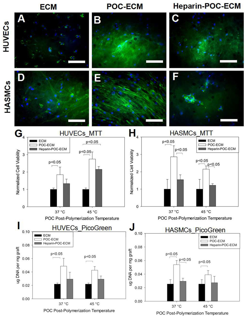
HUVECs (A, B, C) and HASMCs (D, E, F) adhesion onto decellularized aorta ECM (A, D), POC-ECM (B, E) and heparin-POC-ECM (C, F). Green fluorescence (phalloidin) stained for actin, while blue fluorescence (Hoechst) stained for cell nuclei. All images were taken for scaffolds processed at 45°C. Cell viability was analyzed via MTT assay for HUVECs (G) and HASMCs (H) on decellularized aortas with varying modification conditions. In addition, cell adhesion was quantified via PicoGreen assay for HUVECs (I) and HASMCs (J). Scale bar=100µm.

