Figure 2.

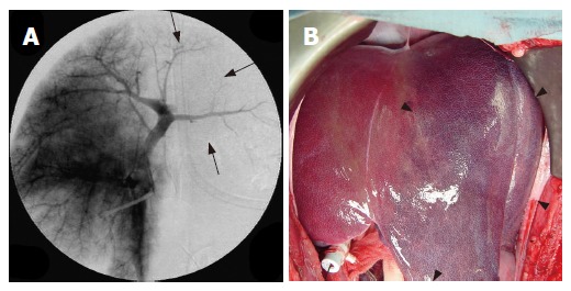
Portography revealing a decrease of portal venous blood flow in the left lobe after LP-TAE before RFA (arrow) (A) and photograph of liver showing the dark red left lobe after LP-TAE before RFA (arrow head) (B).

Portography revealing a decrease of portal venous blood flow in the left lobe after LP-TAE before RFA (arrow) (A) and photograph of liver showing the dark red left lobe after LP-TAE before RFA (arrow head) (B).