. 2015 Mar;31(1):33–40. doi: 10.5487/TR.2015.31.1.033

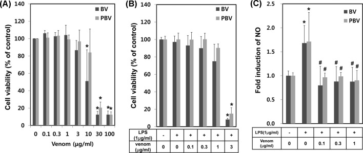
Fig. 2. Comparison of anti-inflammatory effects of BV and PBV. (A) Various concentrations of BV and PBV were applied to macrophage RAW264.7 cells. Briefly, RAW264.7 cells were plated on 24-well and treated with BV and PBV. At 24 hr time period after the treatment, cytotoxicity was measured using MTT assay. (B) RAW264.7 cells were pretreated with LPS for 1 hr and then treated with various concentrations of BV or PBV for additional 24 hr, followed by cell viability assessment. (C) The anti-inflammatory effects of BV and PBV on RAW264.7 were evaluated. At 24 hr time period after the treatment of LPS (1 μg/mL), NO induction level was detected by Griess solution. The data shown are the means ± SD of three independent experiments. Significant difference from control group, *p < 0.05. Significant difference from LPS control group, #p < 0.05.

Fig. 2.