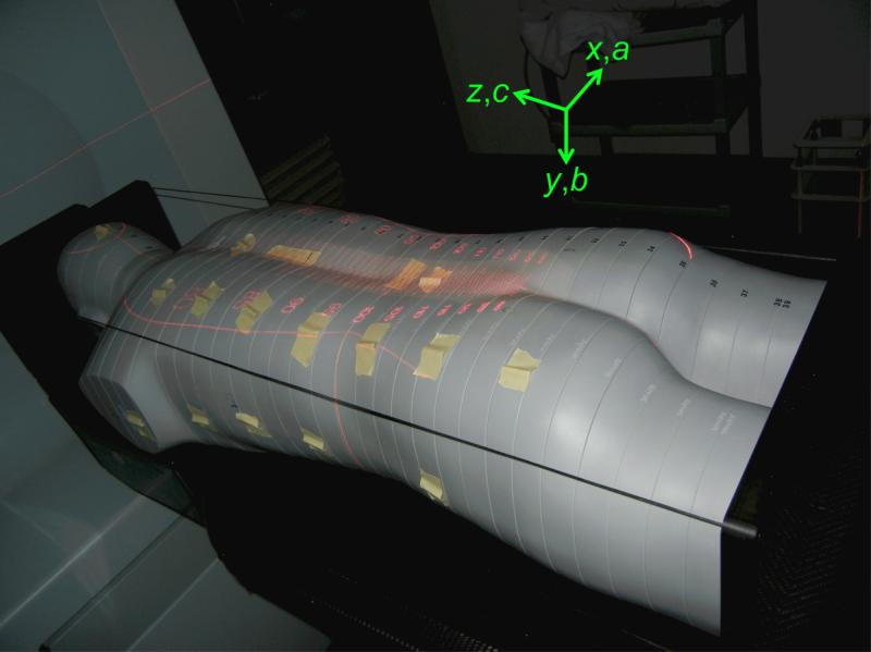
Figure 1.
Photograph of the anthropomorphic phantom placed in the treatment position at AUBMC for the lower spinal field with indicators verifying the source-to-skin distance and the multileaf collimator leaves. TLDs were placed in plug holes throughout the phantom, and they were also taped to the surface of the phantom to measure skin dose in these locations. The axes show the directions of each coordinate in the DICOM patient coordinate system.

