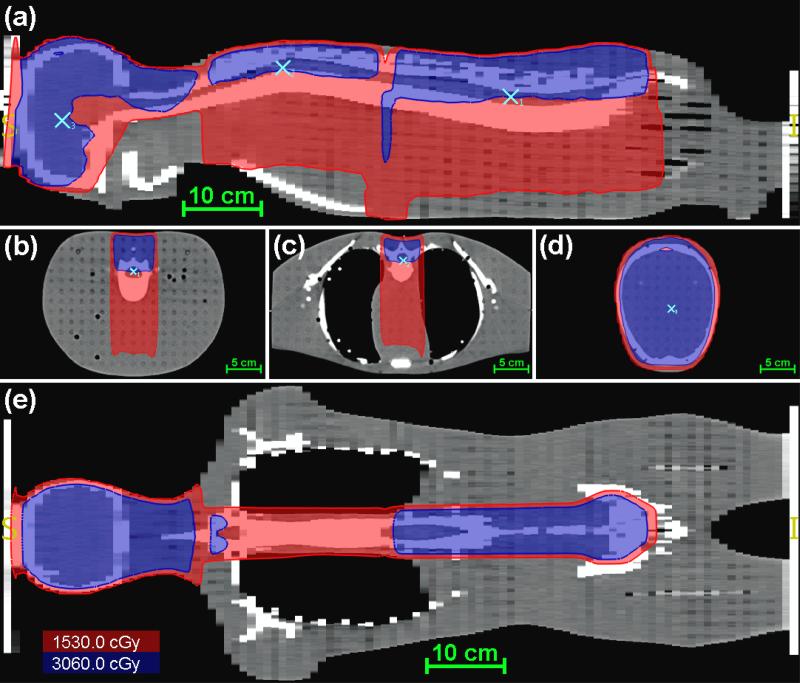
Figure 3.
Dose distribution from the AUBMC TPS for the CSI of an anthropomorphic phantom in the mid-sagittal plane (a), axial planes of the calculation points (b-d), and a coronal plane (e). The red isofill represents the region within the field edge (D ≥ 0.5DRx), the blue isofill represents the region receiving the prescribed dose (D ≥ DRx), and the light blue markers indicate the locations of the calculation points.

