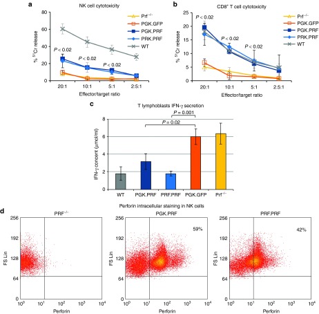
Figure 4.
Lentiviral vector–mediated HSC perforin gene transfer restores T and NK cell cytotoxic function and reduces IFN-γ secretion by T lymphoblasts in vitro. (a) 51Cr release from RMA-S cells co-incubated with NK cells from mice reconstituted with LSK cells transduced with PGK.GFP, PGK.PRF, and PRF.PRF, prf−/− and WT mice. (b) 51Cr release from anti-CD3 bound P815 cells co-incubated with CD8+ T cells from mice reconstituted with LSK cells transduced with PGK.GFP, PGK.PRF, and PRF.PRF, prf−/− and WT mice. The P values correspond to both the comparisons between the PGK.PRF and the PRF.PRF groups with the PGK.GFP group. The highest P value is shown for each point. (c) IFN-γ production by CD8+ lymphoblasts derived from prf−/− mice reconstituted with LSK cells transduced with PGK.GFP, PGK.PRF, and PRF.PRF, from prf−/− and from WT mice after co-incubation with anti-CD3–bound P815 cells for 4 hours. For the three assays n = 3 for each group, and the error bars represent the SD. (d) Representative flow cytometry dot plots for perforin expression in NK cells from one prf−/− mouse, one mouse reconstituted with PGK.PRF, and one mouse reconstituted with PRF.PRF.

